Association of Insulin Resistance With Cerebral Glucose Uptake in Late Middle-Aged Adults at Risk for Alzheimer’s Disease

1
ByCrossFitApril 5, 2020

Question
Does insulin resistance specifically increase risk of Alzheimer’s disease and/or neurodegeneration? If so, how?
Takeaway
This study found increased insulin resistance was associated with lower glucose metabolism in the brain, specifically in regions associated with memory function, and this reduced metabolism was associated with memory impairment. These results are consistent with a larger body of evidence indicating insulin resistance (as seen in diabetes and the metabolic syndrome) is at minimum a risk factor for Alzheimer’s disease and may play a direct causal role.

The ADA estimates more than 29 million Americans, and more than half of those over age 64, are diabetic or prediabetic (1). Insulin action, in both the peripheral and central nervous systems, stimulates glucose uptake and utilization and promotes microvascular blood flow. Previous human and animal research has shown impaired insulin signaling in the brain is associated with brain atrophy, mitochondrial dysfunction, inflammation, memory deficits, reduced cerebral blood flow, and cognitive deficits (2).

As previously discussed on CrossFit.com, these deficits may be related to deficient cerebral glucose metabolism. Impairments in brain glucose metabolism, particularly in regions of the brain associated with memory and executive function, are a consistent feature associated with mild cognitive impairment (MCI) and Alzheimer’s disease (AD); the same impairment is also seen in individuals with the APOE e4 allele (a major genetic risk factor for AD) even when healthy (3).

This observational study sought to understand the link between peripheral insulin resistance — the type seen in prediabetic and diabetic individuals — and reduced brain glucose metabolism and memory function. One hundred fifty middle-aged (age 40-65) adults were recruited from the Wisconsin Registry for Alzheimer’s Prevention. All were cognitively normal. Each subject was assessed for insulin resistance, brain glucose metabolism (via FDG-PET), and memory function.

Higher HOMA-IR levels — i.e., higher levels of insulin resistance — were associated with reduced glucose metabolism throughout the brain (Figure 1). The greatest suppression of glucose metabolism was seen in the medial temporal lobe (MTL) and hippocampus, areas linked to memory function. The degree of insulin resistance was proportional to the degree to which glucose metabolism was suppressed. These two factors were additionally associated with lower scores in various tests of memory function.

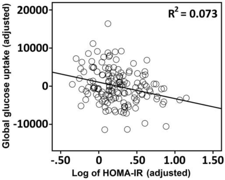
Figure 1: The association between HOMA-IR and global FDG-PET uptake in 150 late middle-aged adults.

The results of the study are consistent with previous work showing peripheral insulin resistance, as well as elevated fasting glucose and insulin levels, are associated with impaired brain glucose metabolism (4). The observed link between insulin resistance, decreased glucose metabolism, and impaired memory provides a potential mechanism for the observed associations between other forms of metabolic disease and Alzheimer’s and supports further efforts to explore this hypothesis (5). As previously reviewed on CrossFit.com, and as noted by the authors of this study, insulin resistance may impair cognitive performance through other mechanisms not directly related to glucose metabolism, including decreased mitochondrial function, oxidative stress, inflammation, and dysregulated lipid metabolism (6).


Notes

  1. Estimates of diabetes and its burden in the United States, 2017
  2. Reduced glucose tolerance is associated with poor memory performance and hippocampal atrophy among normal elderly; Association of metabolic dysregulation with volumetric brain magnetic resonance imaging and cognitive markers of subclinical brain aging in middle-aged adults: The Framingham Offspring Study; Insulin resistance and hippocampal volume in women at risk for Alzheimer’s disease; Insulin resistance and hippocampal volume in women at risk for Alzheimer’s disease; Insulin resistance, brain atrophy and cognitive performance in late middle-aged adults; Low cerebral blood flow is associated with lower memory function in metabolic syndrome
  3. Insulin resistance predicts medial temporal hypermetabolism in MCI conversion to Alzheimer’s disease; Amyloid vs FDG-PET in the differential diagnosis of AD and FTLD; Longitudinal PET evaluation of cerebral metabolic decline in dementia: A potential outcome measure in Alzheimer’s Disease treatment studies; FDG PET and differential diagnosis of dementia; Discrimination between Alzheimer dementia and controls by automated analysis of multicenter FDG PET; Reduced hippocampal metabolism in MCI and AD: Automated FDG-PET image analysis
  4. Higher serum glucose levels are associated with cerebral hypometabolism in Alzheimer regions; Diabetes and elevated hemoglobin A1c levels are associated with brain hypometabolism but not amyloid accumulation; Localization and characterization of insulin-like growth factor receptors in rat brain and pituitary gland using in vitro autoradiography and computerized densitometry; Autoradiographic localization of insulin receptors in rat brain: Prominence in olfactory and limbic areas; Characterizing Alzheimer’s disease using a hypometabolic convergence index; Positron emission tomography imaging and clinical progression in relation to molecular pathology in the first Pittsburgh Compound B positron emission tomography patient with Alzheimer’s disease
  5. Distribution of insulin receptor-like immunoreactivity in the rat forebrain; Metabolic syndrome and cognitive decline; Midlife cardiovascular risk factors and risk of dementia in late life; Meta-analysis of Alzheimer’s disease risk with obesity, diabetes and related disorders; Midlife vascular risk factors and their association with dementia deaths: Results from a Norwegian prospective study followed up for 35 years
  6. Mitochondrial dysfunction is a trigger of Alzheimer’s disease pathophysiology; A “mitochondrial cascade hypothesis” for sporadic Alzheimer’s disease; Role of mitochondrial dysfunction in insulin resistance; Insulin and Alzheimer’s disease: Untangling the web

Comments on Association of Insulin Resistance With Cerebral Glucose Uptake in Late Middle-Aged Adults at Risk for Alzheimer’s Disease

1 Comments

Comment thread URL copied!
Back to 200406
Richard Feinman
April 6th, 2020 at 1:36 am
Commented on: Association of Insulin Resistance With Cerebral Glucose Uptake in Late Middle-Aged Adults at Risk for Alzheimer’s Disease

I removed my previous comment which was an off-the-cuff (translation: wise-ass) response to the figures. The figures are, in fact, not consistent with the conclusion that "HOMA-IR associations with regional glucose metabolism in this study were robust," The figures show a poor correlation by the "eye-ball test," that is, the scatter of the points tells you that, as an end-user,you would not want to make any kind of prediction. The statistics do not conflict with this intuitive response, The indicators of correlation (e.g., β=−0.29) are not strong and an R^2 = 0.178 found for the medial temporal lobe cannot be considered "especially robust." Again, just looking at the figure, the expectation of improving things by changing HOMA is absolutely minimal.


This is different from saying that the conclusion is unreasonable but, in fact, that is a reason for asking for stronger data -- we don't want to repress further research. The authors' disclaimer that the "study was cross-sectional in nature, and no causal inferences about insulin resistance may be inferred" is not in keeping with the description of the results.

(edited)
Comment URL copied!