The flow of blood through the heart follows the same general pattern as the electrical conductive pathway if we begin tracing it from its entry into the right ventricle. However, the blood flow system includes both inflow and outflow. The atrium on the right side of the heart receives deoxygenated blood from the body. This blood is delivered by the superior and inferior vena cava. Blood flow returning from the heart’s innate circulation collects in the coronary sinus and then passes into the atrium through the semicircular Thebesian valve into the atrium.
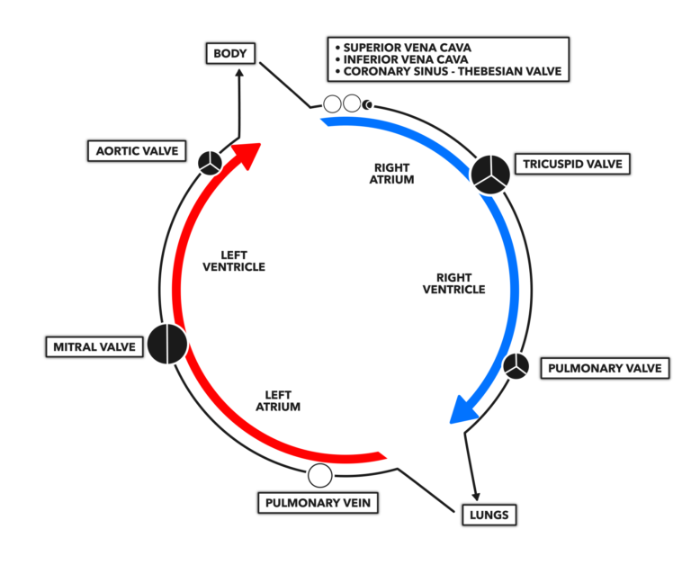
Blood flows through the heart in a cyclical pattern: right atrium → right ventricle → left atrium → left ventricle → and repeat ad nauseum. White circles indicate where no valve is present. Black circles indicate the presence of a valve, with white lines indicating the morphology of the valve.
As atrial pressure increases, the Thebesian valve closes to prevent regurgitation (backward blood flow), and the blood is pushed through the tricuspid valve into the right ventricle. The tricuspid can be identified by the three (tri- = three) leaflets of which it is constructed. Blood ejection from the right ventricle occurs through the pulmonary valve after intra-ventricular pressure pushes it open.
Blood from the right ventricle is oxygenated in the lungs via the pulmonary artery. Once the blood clears the lungs, it returns to the heart by way of the pulmonary vein. It is worthwhile to note that the pulmonary artery and pulmonary vein are exceptions to the rule that arteries carry oxygenated blood away from the heart and veins carry deoxygenated blood to the heart; they have opposite roles to the naming convention.
Oxygenated blood enters directly into the left atrium. Once full, the pressure within the left atrium forces open the mitral valve and blood moves into the left ventricle. The mitral valve has two leaflets, which is why it is sometimes referred to as the bicuspid valve. Rising pressure in the full ventricle shuts the entry valve, opens the aortic valve, and the blood leaves the heart through the aorta.
The aorta is the most important artery in the body in terms of blood delivery. After emerging from the heart, it arches back toward the vertebral column and becomes known as the thoracic then abdominal aorta as it passes through those cavities. All the important appendicular arteries (like the subclavian and iliac arteries) and visceral arteries (like the hepatic and mesenteric arteries) are derived from the aorta. The majority of the blood leaving through the aorta is destined for systemic circulation, but a portion of it is immediately redirected from the aorta into coronary circulation.