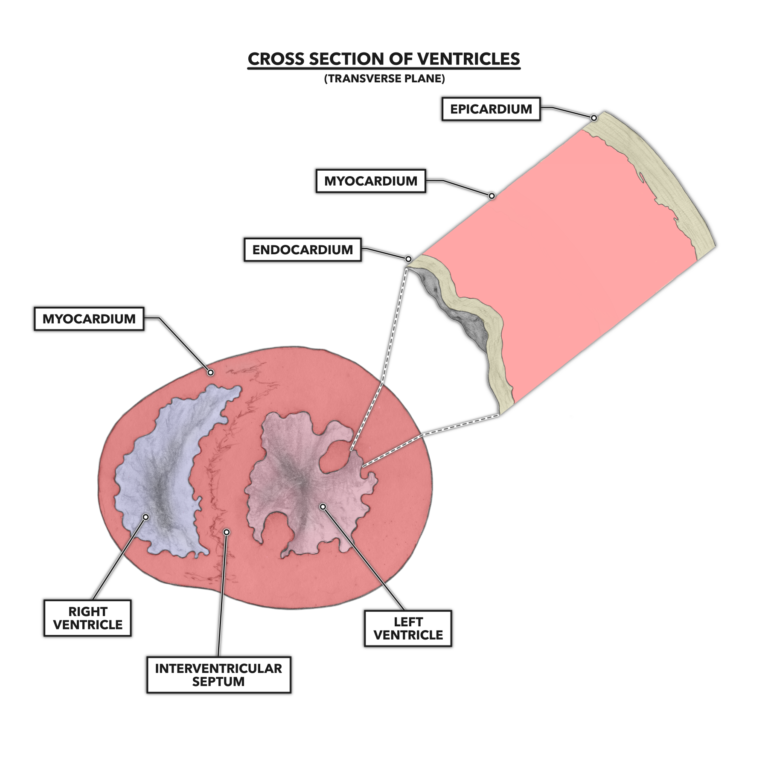
The wall of the heart is composed of three distinct layers: the epicardium, myocardium, and endocardium. The epicardium is the outermost layer and is in close communication with the inner layer of the pericardium. The endocardium is the innermost layer and is fairly thin but important. It contains the innermost lining of the heart chambers, a single-cell layer called the endothelium. The endothelium produces and secretes a large number of bioactive chemicals important for circulatory function. The myocardium is the middle layer between the epicardium and endocardium. The Latin prefix “myo-” indicates this layer contains muscle. It also contains the bulk of the ventricular walls and septum.
The muscle present in the heart differs anatomically and functionally from skeletal muscle. Cardiomyocytes (myocyte = muscle cell) differ from skeletal myocytes in the following four ways:
- Morphology – Cardiomyocytes have a more jigsaw puzzle-like appearance; they are not fusiform (i.e., tapering at both ends) and are smaller than skeletal muscle cells.
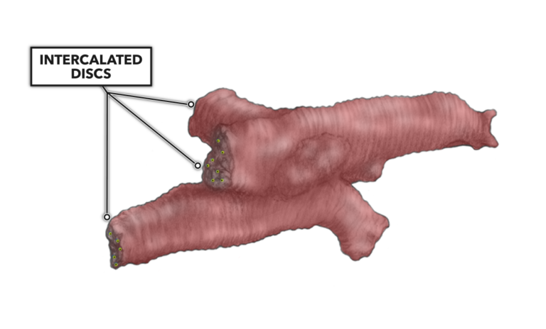
- Intracellular communication – Many intercalated discs are present in cardiac myocytes. These intercalated discs connect individual heart muscle cells so they can function together as an organ.
- Nucleation – Cardiomyocytes are mononucleated rather than multinucleated.
- Contractility – Cardiomyocytes are autorhythmic, meaning they can spontaneously contract without neural input.
The unique morphology of cardiomyocytes increases the heart’s communicative surface area. Communication between cells occurs through the intercalated discs. There are also little pores between two adjacent cells called gap junctions (tiny yellow dots below) that allow free and rapid passage of chemical signals between cells. This enables simultaneous muscular contraction of a segment of the heart. For example, when a cell or set of atrial muscle cells is stimulated to contract, the communicative signal cascade made possible by these features drives all atrial muscle cells to contract. The same action occurs later in the ventricles.
The actions of the atrial and ventricular segments are sequential: The atria contracts first, then the ventricles. Such sequencing is made possible by the presence of specialized electroconductive cells.
Comments on The Heart, Part 3: Muscular Composition and Arrangement
0 Comments