Many muscles in the thoracic segment contribute to respiratory function by moving the costals. Examples of this are the external and internal intercostals, which are found circumferentially around the thoracic cavity — posterior, lateral, and anterior. The transversus thoracis and diaphragm also contribute to respiration.
Transversus thoracis (Figure 1) – The transversus thoracis muscles are bilateral, thin multi-segment muscles fanning laterally and upward from the xiphoid process on the front of the chest wall. From its inferior attachment to the sternum, each segment attaches to the second through sixth ribs (costals). The attachment to the sixth costal has a horizontal fiber orientation. As the attachments ascend to the second costal, the angle of fiber orientation nears vertical. This has implications for function.
The overall function of the muscle pair is to aid in forced expiration. Specifically, the sixth costal segment, owing to its horizontal orientation, stabilizes the sixth rib and allows the upper four segments to pull the second through fifth ribs down. This motion decreases the diameter and height of the rib cage.
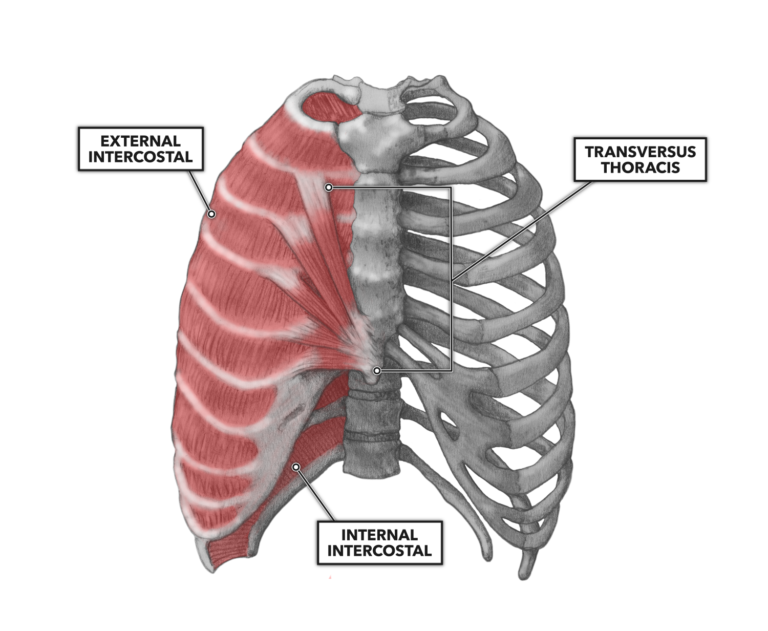
Figure 1: The transversus thoracis muscles are bilateral, thin multi-segment muscles fanning laterally and upward from the xiphoid process on the front of the chest wall.
Diaphragm (Figure 2) – The diaphragm is a muscle with a large surface area that spans the inner diameter of the inferior thoracic cavity (the chest). Structurally, it demarcates the division of the thoracic cavity above and abdominal cavity below. Functionally, it is the most important muscle contributing to respiration (breathing).
The muscle attaches to the inner and lower aspects of the sixth through 12th ribs, the inner xiphoid process, the 12th thoracic vertebra, and the first and second lumbar vertebrae. From these outer attachments, the muscle fibers curve upward and inward to merge into a large central tendon. The muscle and central tendon create a domed structure that rises within the thoracic cavity.
Contraction of the diaphragm pulls the central tendon down, expanding thoracic volume. This creates negative pressure within the lungs (a vacuum), which drives inward airflow. Relaxation of the muscle allows the central tendon to rise back to its higher original position, decreasing thoracic volume. The resulting positive pressure within the lungs expels air.
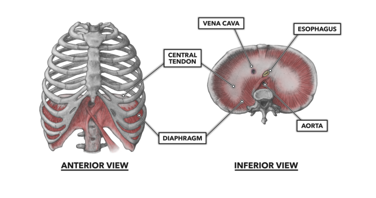
Figure 2: The diaphragm is a muscle with a large surface area that spans the inner diameter of the inferior thoracic cavity (the chest).