As we progress down the vertebral column, the next 12 vertebral bones below the last cervical vertebra are thoracic vertebrae. The thoracic vertebrae, along with the breastplate, thoracic cavity, and thoracic wall, make up the thorax. These vertebrae are easy to identify, as they articulate with the ribs. The first thoracic vertebra articulates with the first pair of ribs, the second thoracic vertebra articulates with the second pair of ribs, and so on, down through the 12th thoracic vertebra and 12th pair of ribs. The accepted abbreviations for the thoracic vertebrae are T1 through T12. On most individuals, you can palpate the spinous processes on all 12 vertebrae.
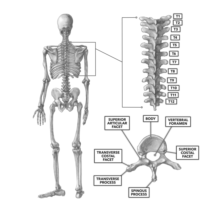
Figure 1
What sets the structure of the thoracic vertebrae apart from the cervical and lumbar vertebrae is the presence of costal facets, the articulation points for the costal bones (ribs). Each thoracic vertebra has six individual costal facets where it articulates with costal bones. One set, the pair of superior articular facets, is made up of shallow depressions on the posterior and lateral portion of the vertebral body. The second set, the pair of transverse costal facets, is made up of shallow depressions on the anterior lateral surface of the transverse processes. A third set is found directly below the superior costal facets on the anterior inferior surface of the vertebral body (not shown). Combined, these facets articulate with the ribs to keep their posterior ends seated upon the vertebral column.