The vertebral column—also referred to as the spine, spinal column, or backbone—is the medial and vertical axis of the body. The major component of the axial skeleton, it is comprised of 33 bones, each of which interlocks with the bones immediately above and below. The bones of the vertebral column can be divided into five groups (Figure 1):
- The cervical vertebrae are the smallest and most superior of the vertebrae. The most superior of these vertebrae articulate with the skull.
- The thoracic vertebrae are matched to each pair of ribs to form the rib cage and establish the thoracic cavity.
- The lumbar vertebrae are the largest and thickest of the vertebrae and carry the largest load of the moveable vertebrae. The last lumbar vertebrae articulate with the sacrum below.
- The sacrum forms the base of the spine and is constructed from five fused vertebrae.
- At the very inferior tip of the sacrum is the coccyx. It is formed from four or fewer fused vertebral bones.
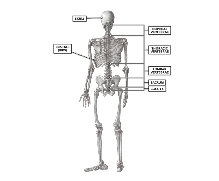
Figure 1. The bones of the vertebral column can be divided into five groups: the cervical vertebrae, thoracic vertebrae, lumbar vertebrae, sacrum, and coccyx.
The bodies of vertebrae become thinner the higher they are on the vertebral column. Vertebral bodies are separated from each other by intervertebral discs made of tough fibrocartilage. Each individual vertebra, from the first cervical vertebra down to the last lumbar vertebra, is relatively unique in size and shape, owing to its anatomical position and function along the vertebral column. All prototypical vertebra, however, may be divided into three basic parts (Figure 2):
- An anterior rounded chunk of bone called the body. This part of the bone carries the weight of the body above it.
- A group of posterior projections and processes that serve as articular surfaces for adjacent vertebrae and sites of attachment for ligaments and muscles.
- A large central hole called the vertebral foramen. The sequential stacking of the vertebrae on top of each other provides a canal through which the medulla spinalis (spinal cord) passes from its lower terminations up to its passage into the cranium and the brain.
NOTE: Vertebra is singular and vertebrae is plural.
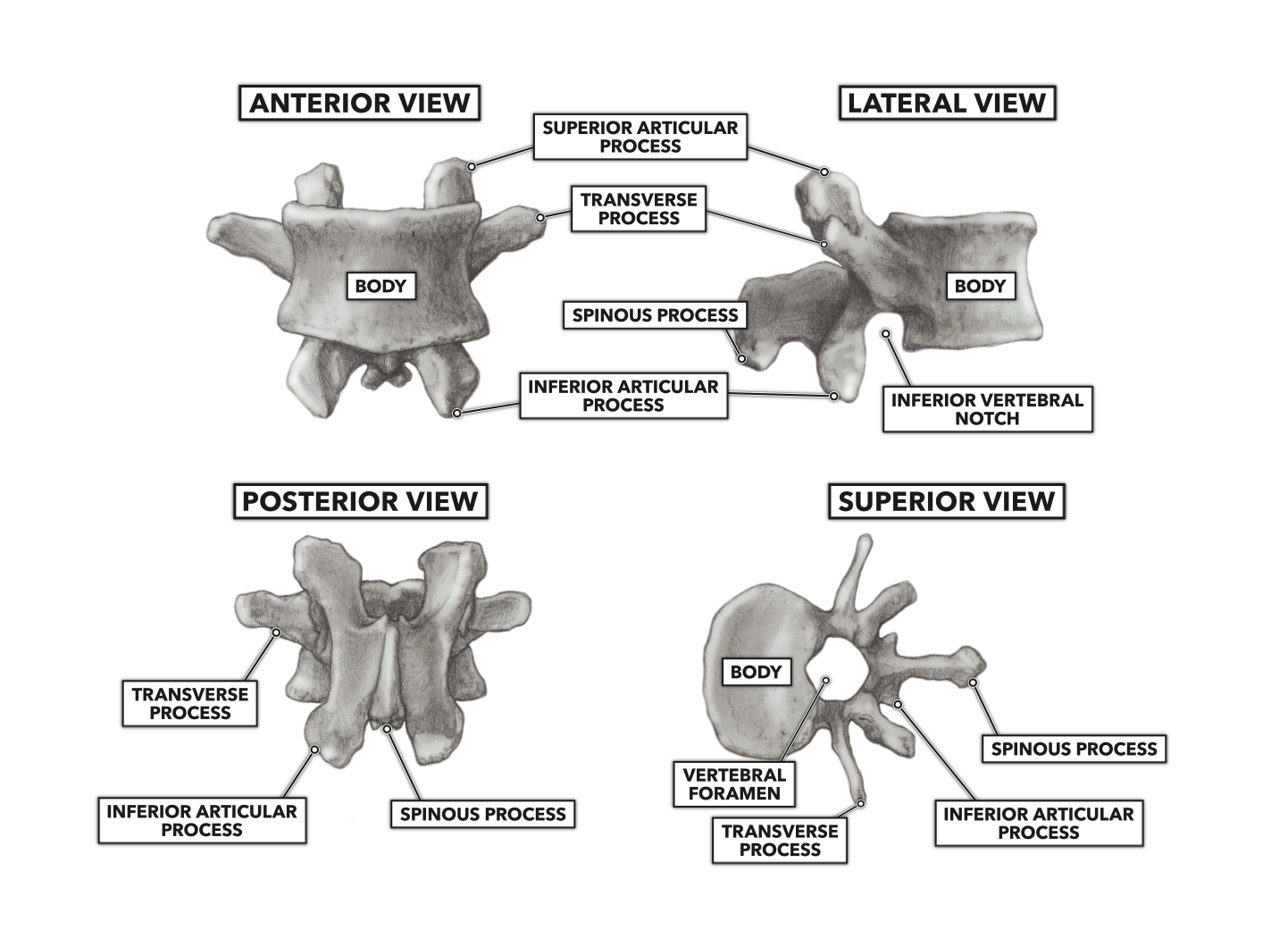
Figure 2. A prototypical vertebra is divided into three basic parts: the body, a group of posterior projections and processes, and the vertebral foramen.
The superior and inferior surfaces of a vertebra are not flat; rather, they have shallow saucer-like concavities. The deepest part of the concavity is toward the anterior, and the shallowest part is to the posterior. Within the space on both the superior and inferior body surfaces, the concavity is lined with a robust hyaline cartilage endplate, upon which a tough but somewhat pliable intervertebral disc lies (Figure 3a).
The intervertebral disc is comprised of two parts (Figure 3b):
- The annulus fibrosus is an interlocking and concentric set of collagenous fiber rings. The rings are thicker toward the outer edges and thinner toward the center. This structure aids in containing nucleus pulposus and distributing hydraulic forces across the intervertebral joint under load.
- The nucleus pulposus is a gel-like substance packed within a collagenous sac surrounded by the annulus fibrosus. The contents of the sac are suspended cells and mucoproteins such as collagen, aggrecan, hyaluronan, chondroitin sulfate, and keratin sulfate.
Combined, the vertebrae, endplate, and intervertebral disc structure protect the medulla spinalis and allow a small degree of individual intervertebral movement that, when accomplished in concert with the entire vertebral column, can produce the large movements of the trunk we use in training and everyday tasks.
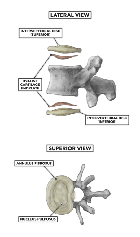
Figure 3 a & b. Components of the intervertebral discs