The muscular structure of the wrist and hand is quite complex, reflecting the complexity of the tasks they perform. The high degree of joint mobility required for manual dexterity involves many muscles that can be precisely controlled. The human wrist and hand fulfill this requirement with 20 sets of muscles acting on the joints to produce movement. Muscles that contribute to wrist control go high enough up the system to require shoulder attachments. For example, the biceps brachii can act to rotate the radius and therefore the hand and wrist. The majority of wrist flexors, extensors, and rotators attach proximally to the humerus, radius, and ulna and attach at various distal sites, including the carpals, metacarpals, and phalanges.
We often see the muscles of the wrist categorized by function (i.e., as a flexor or extensor) and location (anterior or posterior). Such characterizations are fairly apt, as flexors are found on the anterior side of the arm and extensors are found on the posterior. We will see a few exceptions to this, however, where anterior muscles carry out an action other than flexion.
SUPERFICIAL ANTERIOR FLEXORS
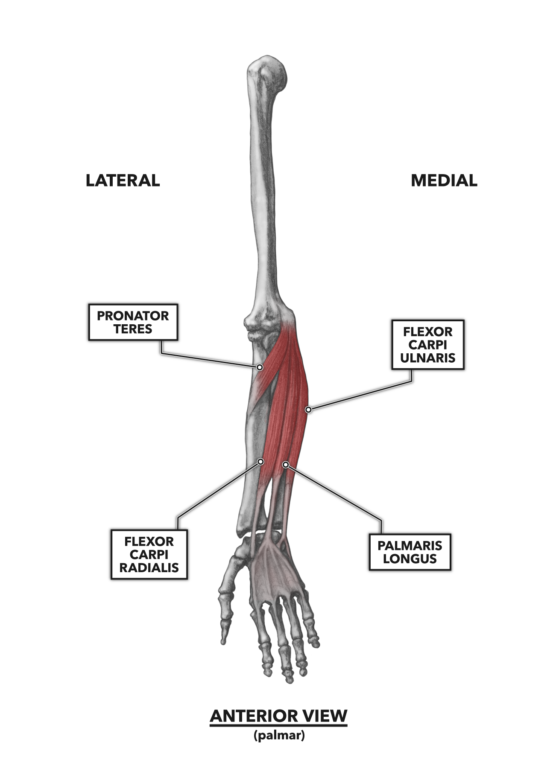
Figure 1: The most superficial of the wrist flexors are the flexor carpi radialis, palmaris longus, flexor carpi ulnaris, and pronator teres. The orientation of the pronator teres is a short angle from the medial epicondyle of the humerus and the medial ulna down to the lateral radius. This makes it a strong pronator and an exception to the anterior-posterior/flexor-extensor rule.
Flexor carpi radialis – The name of this muscle provides all you need to know. It is a flexor, and since flexors are anterior, it must be on the anterior side of the arm near the radius. It attaches distally to the second and third carpi (“carpi” is the plural of “carpal”). It is attached proximally to the medial epicondyle of the humerus. The basic actions driven by contraction of the muscle are flexion of the hand and, as the muscle angles across the forearm, medial to lateral, it assists in abduction of the hand. The muscle is also a weaker elbow flexor.
Palmaris longus – This muscle attaches proximally to the medial epicondyle of the humerus and distally to the proximal center of the palmar aponeurosis, a fanned sheet of fascia lying across the palm into which many ligaments and tendons invest themselves. The tendon running between the muscle and the aponeurosis is quite long. When the muscle contracts, the palmar fascia is tensed. This is a small muscle that may actually be absent in 10% or more of the population. It has been reported in the clinical literature that its absence does not affect wrist flexion or grip strength — an interesting finding given that flexion of the wrist is its primary function. The muscle is also a weaker elbow flexor.
Flexor carpi ulnaris – This muscle can be found along the medial side of the palmaris longus, forming the contour of the medial forearm. The “flexor” in its name indicates both its function and location on the anterior side of the forearm, “carpi” indicates it has an attachment to at least one carpal, and “ulnaris” suggests it is a muscle associated with the ulna. The flexor carpi ulnaris attaches proximally to the medial side of the olecranon, but it also has a proximal attachment to the lower aspect of the medial epicondyle. It attaches distally to the anterior surface of the pisiform bone. The muscle’s anterior location makes it a flexor; its medial attachments also make it an adductor of the hand. The muscle is also a weaker elbow flexor.
SUPERFICIAL ANTERIOR PRONATOR
Pronator teres – This muscle angles across the upper forearm. It has proximal attachments at the supracondylar ridge of the humerus and medial aspect of the proximal ulna. It then angles down, superior to inferior, to attach to the lateral aspect of the radius, about one-third down its length. It is this angular orientation, medial to lateral, that makes it a strong pronator. The muscle attaches to three bones: the radius, humerus, and ulna. The radius is situationally more mobile than the humerus, and as the ulna is seated into the olecranon fossa of the humerus, ulnar rotation is prevented. Thus, the action of the pronator teres is to pull the lateral aspect of the radius up and over medially (medial rotation, internal rotation, or pronation). The muscle is also a weaker elbow flexor.
INTERMEDIATE ANTERIOR FLEXORS
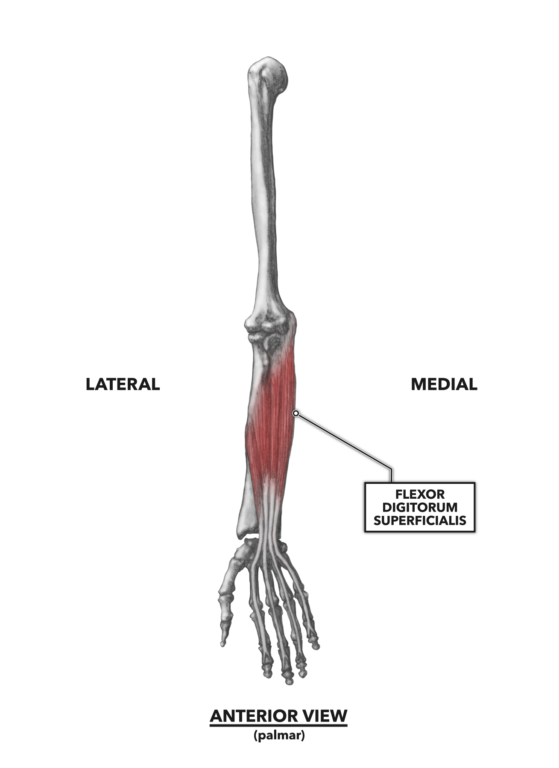
Figure 2: The flexor digitorum superficialis can be found lying beneath the superficial flexors. Only portions of this muscle are actually superficial.
Flexor digitorum superficialis – This is a finger flexor, as the name (digitorum) suggests. It lies beneath the brachioradialis, pronator teres, flexor carpi radialis, and palmaris longus. The flexor digitorum longus proximally attaches at the distal radius and medial epicondyle of the ulna. Distally, it divides into four tendinous splits and attaches to the intermediate phalanx of phalanges two through five. It is a flexor, active in wrist flexion and flexion of the fingers, as in making a fist.
DEEP ANTERIOR FLEXORS
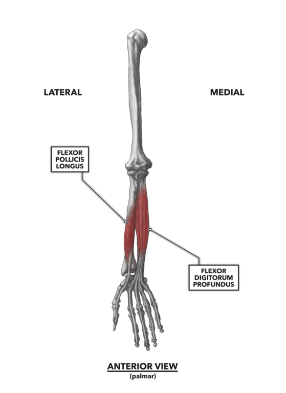
Figure 3: The deep anterior flexors, the flexor pollicis longus and flexor digitorum profundus
Flexor pollicis longus – “Pollux” is the Latin nomenclature for the thumb, so the flexor pollicis longus is a flexor of the thumb. It attaches proximally to the upper half of the radius and includes an attachment to the coronoid process of the ulna. It also attaches distally to the base of the distal phalanx of the thumb. As it crosses the wrist joint, it is also a wrist flexor.
Flexor digitorum profundus – As its name suggests, this muscle is a flexor of the fingers. In Latin, “profundus” can mean “deep,” so this indicates the muscle’s location. It has a long proximal attachment along the ulna, spanning from near the olecranon down about two-thirds its length. The distal attachments of the profundus are interesting and relatively unique. Instead of a single tendon emerging from the muscle and then dividing into slips for multiple attachments, four tendons arise independently along the distal border of the muscle belly. The tendons then attach distally to the bases of the distal phalanx of digits two through five.
DEEP ANTERIOR ROTATOR
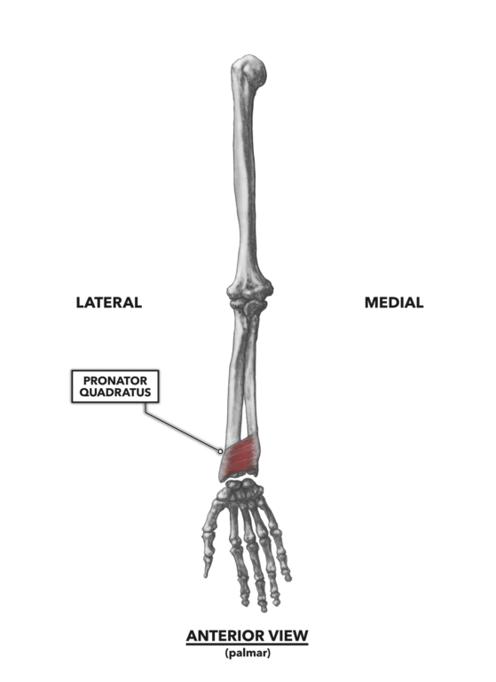
Figure 4: The pronator quadratus is another exception to the anterior-posterior/flexor-extensor rule. It spans the distal radius and ulna only and does not cross the wrist, so it cannot be a flexor.
Pronator quadratus – This is an anterior muscle that is not a flexor. It is, as the name implies, a pronator of the hand. It attaches along the distal aspect of the last few inches of both the radius and ulna, forming a roughly quadrilateral shape. The direction of the fiber orientation is lateral across the space between the two bones. The radius is the most mobile of the two bones, as the ulna is seated in the olecranon fossa and this prevents its rotation. Thus, the action of the pronator quadratus is to pull the lateral aspect of the radius up and over medially (medial rotation, internal rotation, or pronation).
To learn more about human movement and the CrossFit methodology, visit CrossFit Training.