We often think of muscles only in the context of biceps, triceps, pecs, and quads, but to do so ignores the fact that there is more than one type of muscle cell in the human body. There are, in fact, three types of muscle cells:
- Skeletal muscle cells drive movement and shape our physique.
- Cardiac muscle cells create the rhythmic and persistent contractions of the heart to circulate blood.
- Smooth muscle cells, the type found in vascular walls, intestine walls, and the uterus, act slowly create movement along tubular tracts.
Each cell type possesses characteristics that make it particularly well suited for its anatomical location and in its role in contracting or generating force. The most obvious differences between cell types relate to shape and size. Smooth muscle cells are basically fusiform (spindle-shaped) and can be as small as about 20 microns in length (e.g., in arterioles) and up to 500 microns in length (e.g., in the uterine wall). Cardiac muscle cells are generally presented conceptually as two-dimensional jigsaw pieces but are actually quite variant three-dimensional cells of about 85 microns in length. Skeletal muscle cells are approximately fusiform, long and fiber-like. They range in length from 1,000 microns in the stapedius muscle to up to 500,000 microns in the sartorius muscle.
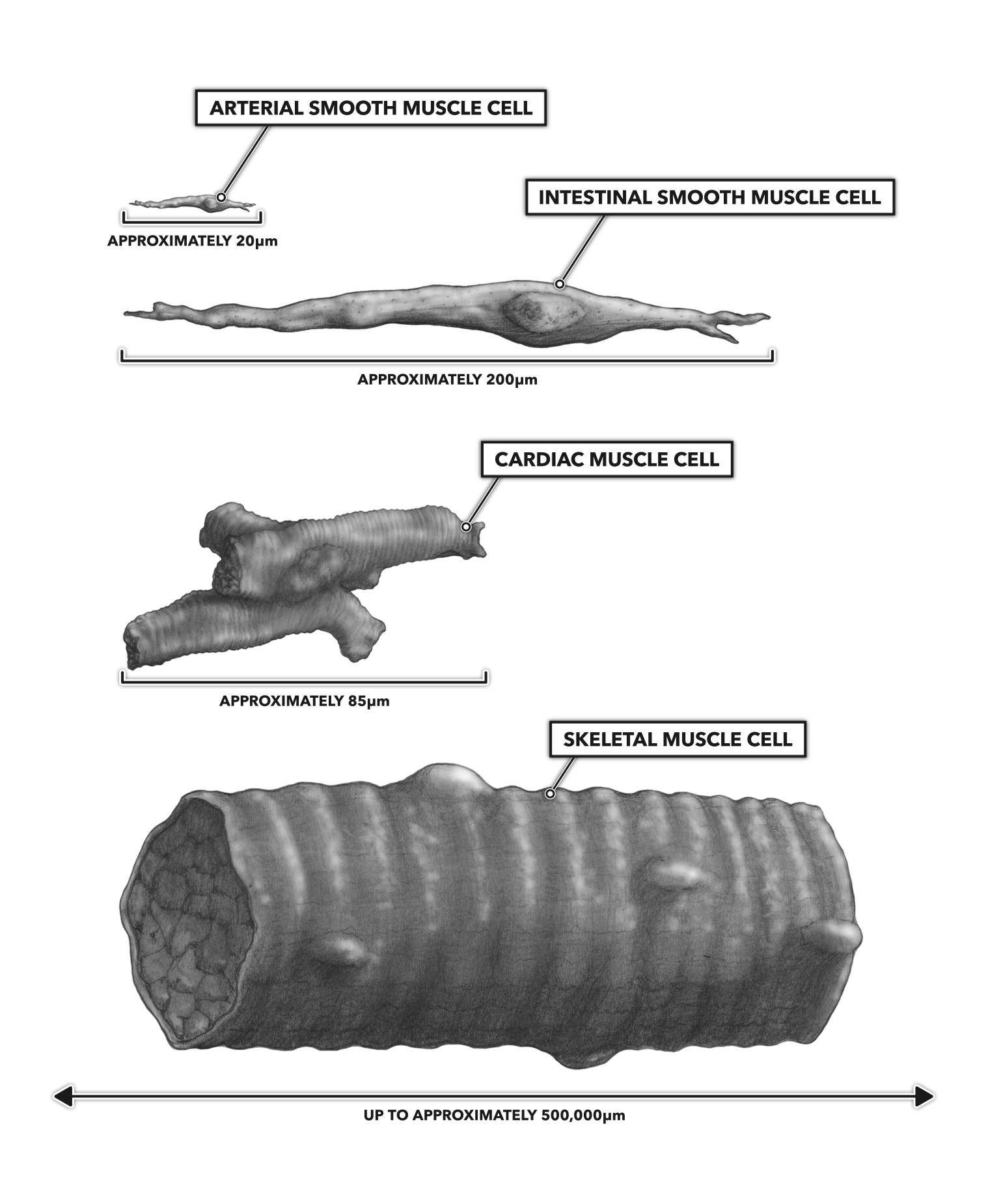
Figure 1: Muscle cell types and dimensional estimates
Another difference is in the cell nuclei: Smooth and cardiac muscle cells possess a single nucleus per cell, and skeletal muscle cells are multinucleated. There are about five nuclei per 100 microns of cell length in skeletal muscle (1).
Within each muscle cell type are a set of chemicals that interact to become biological motors. These chemicals interact, generate force, and if the force is great enough, create movement. At the heart of these micro-motors are two contractile proteins called actin and myosin. These myofilaments are differentially arranged in the cell types. In smooth muscle, the filaments are distributed as a mesh throughout the cell, and the ends of the filaments are attached to dense bodies associated with the cell membrane.
Specifically in skeletal muscle and to a lesser extent in cardiac muscle, the actin and myosin filaments are arranged in a linear manner that gives the muscle a striated or striped appearance. This results from the physical structures of myosin, a very large and thick protein with a globular end, and actin, a thinner more linear protein. Myosin is a dimer, formed of two individual myosin proteins bound together. Actin is a polymer, formed of multiple actin molecules strung together along a chain. The two sets of myofilaments, one thick and the other thin, are arranged in alternating lines, separated by a space bisected by a structural protein called myomesin. Myosin is held in place by titin (another specialized protein). Each set of actin myofilaments is bound to another structural protein called alpha actinin. This composite structural and functional unit is known as a sarcomere. The chemical interactions of the thick and thin filaments within the context of the related structural protein framework directly create muscle contraction.
An individual sarcomere is about 2.5 to 3.0 microns long. A myofibril is formed of a sequence of sarcomeres. A single muscle cell from a latissimus dorsi will have about 100,000 sarcomeres oriented in sequence (2). Skeletal muscle fibers are made up of a bundle of myofibrils.
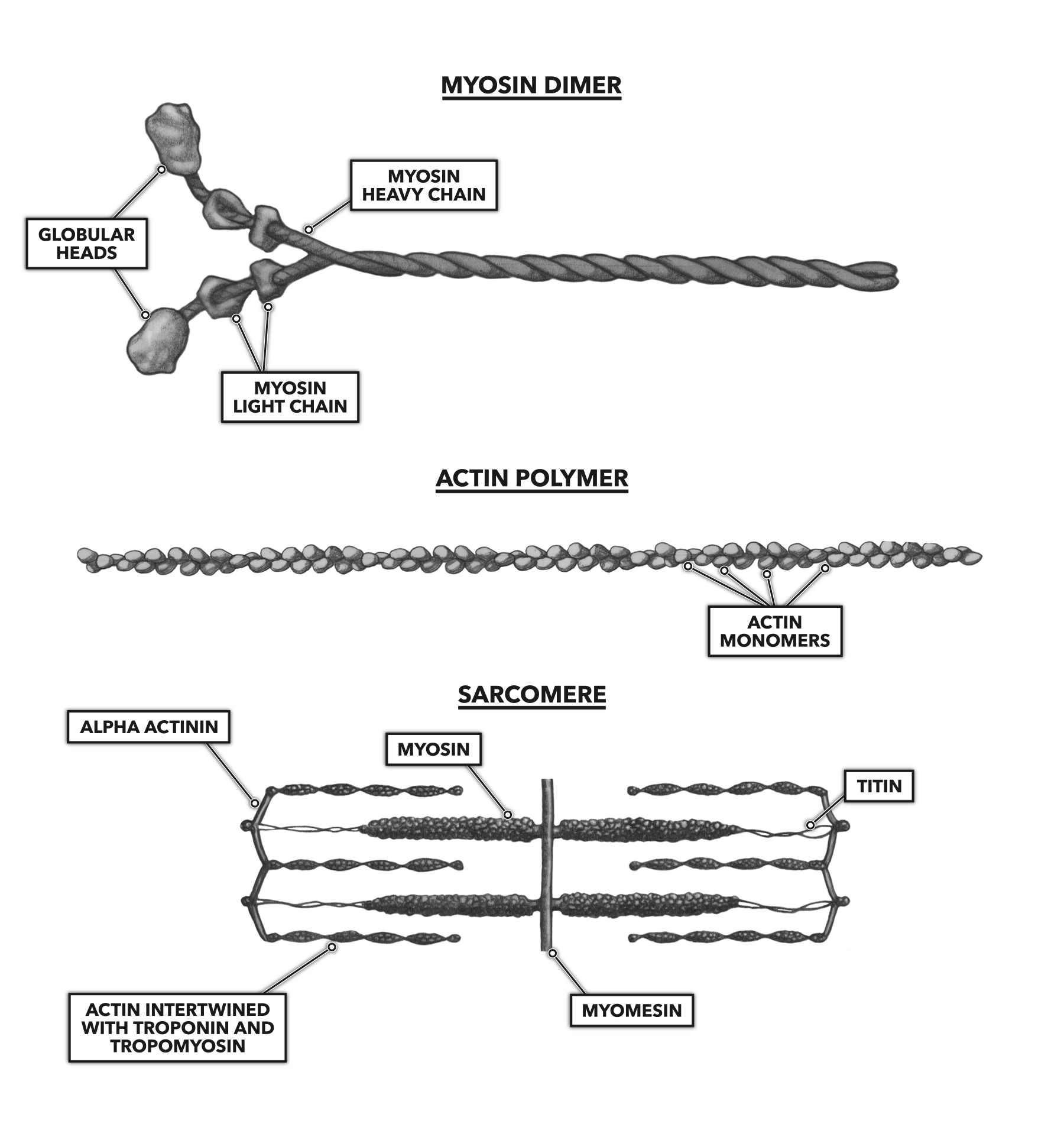
Figure 2: The arrangement of proteins in a sarcomere
References
- Bruusgaard JC, Liestøl K, Ekmark M, Kollstad K and Gundersen K. Number and spatial distribution of nuclei in the muscle fibres of normal mice studied in vivo. Journal of Physiology 551.2(2003):467–478.
- Gerling ME and Brown SH. Architectural analysis and predicted functional capability of the human latissimus dorsi muscle. Journal of Anatomy 223.2(2013):112-22.
To learn more about human movement and the CrossFit methodology, visit CrossFit Training.