Fat, Part 2: Microanatomy

4
ByCrossFitJuly 22, 2020

Fat cells, also known as monovacuolar cells or adipocytes, are essentially large lipid droplets surrounded by a thin cell layer. The cells are mononucleate, with the nucleus found on the periphery of the cell, bundled with other organelles in the largest accumulation of cytoplasm. The remainder of the cytoplasm is spread thinly around the adipocyte cell boundary. This provides an appearance of a cell membrane stretched around the lipid with pockets of cytoplasm appearing randomly on the surface. The lipids stored inside those cells are primarily triglycerides (three fatty acids bound to a glycerol backbone).

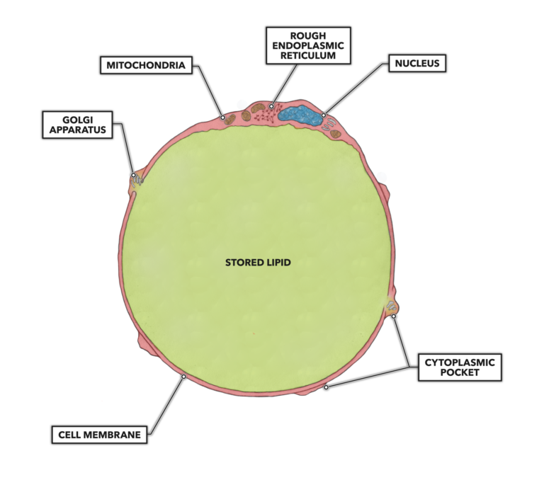
Figure 1: Adipocyte structure

Humans have a large range of cell sizes. Although the average size is frequently reported to be 0.1 millimeters (100 μm), data indicates a much more varied norm, with individuals exhibiting an average cell size of 70 μm and a peak cell size of 110 μm (1). But these dimensions can be much smaller or tremendously larger depending on the nutritional state of the individual. Obese individuals will tend to have larger, hypertrophied fat cells compared to slimmer people.

Fat cells and fat tissue are adaptive entities that can be affected by nutritional habits and exercise. The cells can adapt to store more energy by simply adding more fat to their contents (hypertrophy of fat cells), but there is a critical volume when additional mass cannot be added to individual cells. When about four or more times the normal content of fat is present, it is thought that a size limit is reached and the adipocyte will become mitotically active and begin cell division (hyperplasia of fat cells) to open up new lipid storage space.

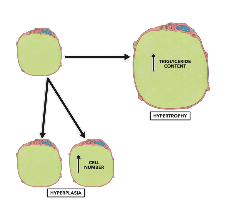
Figure 2: Hyperplasia of fat cells

This latter process, fat cell hyperplasia, occurs in extreme obesity, and most of us are not in danger of adding new fat cells. What we are in danger of is adding new triglycerides to the interior of our existing adipocytes simply by eating a caloric excess, consuming an inappropriate nutritional composition, or by underexerting ourselves and burning too few calories.

In isolation, an adipocyte assumes a spheroid shape, but when collated into fatty tissue, the cells are shaped by pressures from adjacent cells and tissues. If you look at fat cells under a microscope, you will note they look reminiscent of the arrangement of styrofoam beads in the wall of a cooler or like an assembled mass of polyhedral dice.

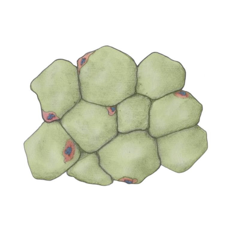
Figure 3: Collated fat cells in fat tissue

In infants and to a much lesser extent in adults, there is a second type of fat tissue called brown fat. Composed of a subtype of adipocytes that are more metabolically active than white fat, brown fat produces heat that helps keep infants warm. These cells are multinucleate and have more cytoplasm, more bioenergetic organelles, and less fat content than white fat cells. In adults, brown fat is largely absent, found in exceedingly small amounts in the neck and shoulders in some humans.

Although adipocytes comprise the major mass of cells present in adipose tissue, fibroblasts, macrophages (leukocyte found in tissue not blood), and endothelial cells are also present as part of the structural unit. As one would expect, there are quite a few blood vessels perfusing fat tissue. This facilitates lipid deposition (adding fat contents to an adipocyte) and lipid mobilization (removing fat from an adipocyte).


Additional Reading


References

  1. McLaughlin T, et al. Subcutaneous adipose cell size and distribution: Relationship to insulin resistance and body fat. Obesity, 22.3(2014): 673–680. 2014.

To learn more about human movement and the CrossFit methodology, visit CrossFit Training.

Comments on Fat, Part 2: Microanatomy

4 Comments

Comment thread URL copied!
Back to 200723
Tyler Hass
July 24th, 2020 at 9:50 pm
Commented on: Fat, Part 2: Microanatomy

I'm interested in the second paragraph on cell sizes. Am I correct that the dimension is the volume? A Google search yields 100um^3 as the average cell size in the human body, but that is substantially influenced by the small size of red blood cells. Adipocytes are listed as 600,000um^3. (source)


Here, I found an article comparing adipocyte size of identical twins. The average adipocyte in the obese co-twins was 547,000 um^3, while for the lean co-twins it was 356,000 um^3. They found in half the pairs, the obese fat cells were the same size as in their lean co-twin, but in larger number. Citing the paper:


“Obese co-twins who had an increased adipocyte count compared with their lean co-twins (hyperplastic obesity) were indistinguishable from their lean counterparts in most metabolic measures (Figure 2). In contrast, obese co-twins who had a more hypertrophic obesity with a decreased cell count compared with their co-twins had significantly more liver fat, insulin resistance, inflammation and LDL cholesterol than the lean twin pair members (Figure 2).”


Very interesting and totally in line with what the article said about hyperplastic vs hypertrophic adipocytes.

Comment URL copied!
jr Wild
July 25th, 2020 at 5:06 am

There is something interesting within dietfits study after all. The referenced ms. Mclaughlin found out, that size matters. Fat cell size. https://www.healio.com/news/endocrinology/20191205/despite-similar-weight-loss-lowcarbohydrate-diet-delivers-insulin-reductions-smaller-fat-cells-than 


Dietfits finding was, that any shift from SAD towards whole foods is beneficial (yawn ), and insulin sensitivity and resistance had less than expected affect to weight. However, those with lower carb diet clearly decreased their fat cell size.


If we consider fat cell as a balloon, LC group increased their intake capacity and available flexibility of storage cells. Call it more insulin sensitivity, for example.


If a fat cell is over filled and starts leaking free fatty acids to blood stream by itself, inappropriately, it isn't only insulin resistance -it is a starting kick for t2d. Talk about personal fat treshold.


As meantioned by Tyler, the size is different within obese and lean individuals. I wonder the beginning of visceral deposits; is it chronic insulin excess (excessive and lengthy excretion counts also), or excess leaked FFA that needs to find a home, or both together?


Looking forward to final report of Dr. Maclaughlin!

JR

Comment URL copied!
Tyler Hass
July 26th, 2020 at 7:59 pm

JR,

That's a great study. Thanks for sharing it.

One factor in insulin resistance is cell membrane fluidity. I've heard insulin receptors don't work as well on a rigid cell membrane. It helps to have a bit of wiggle room in the process of docking insulin to its receptor on the cell membrane.

A variety of factors like the fatty acid composition of the membrane and glycation can make a cell membrane more rigid, which increases its insulin resistance. It's plausible that a cell that has swollen too much much will become more rigid, much like an overfilled balloon. In the overfilled state, it would make sense to block the anabolic effects of insulin. So, insulin resistance could be a homeostatic mechanism to protect the cell from excess hypertrophy.

This paper has some good insights: Revisiting the membrane-centric view of diabetes


"SFAs obtained from the diet or via lipogenesis in the liver and adipocytes pose a relentless challenge to fluidity-sustaining systems, even more so in genetically predisposed individuals. This is likely exacerbated by unnatural fats of various types generated during the production of margarines or superheated vegetable oils used for frying much of our (fast) foods, and which may not be handled efficiently by the cellular machinery. Chronic low fluidity in our membranes has several diabetes-promoting consequences, including impairing insulin secretion and signaling, reduced efficacy of GLUT4 localization to membranes and hardening of blood vessels. The idea that low membrane fluidity is an important component of diabetes pathophysiology is an old one that has been reviewed a few times."

Comment URL copied!
Nathan McFall
July 23rd, 2020 at 8:12 am
Commented on: Fat, Part 2: Microanatomy

Always informative and enlightening to a nutritional layman like myself.

Comment URL copied!